
Nanobody preparation tips sharing
Heavy chain antibodies are special antibodies naturally occurring in camels and cartilaginous fish consisting of only two heavy chains, including a heavy chain variable region (VHH) and two conventional CH2 and CH3 regions, which are naturally missing. Heavy chain antibodies bind antigens through a variable region (VHH) on the heavy chain, which can be stably present alone in vitro and is called camel single-domain antibody (SdAb) or nanobody (Nanobody). The nanobody crystal diameter is 1.2 nm, about 5 nm long, and the molecular weight is only 4/1 (about 10 kD) of the traditional intact antibody, but it still has complete antigen recognition ability.
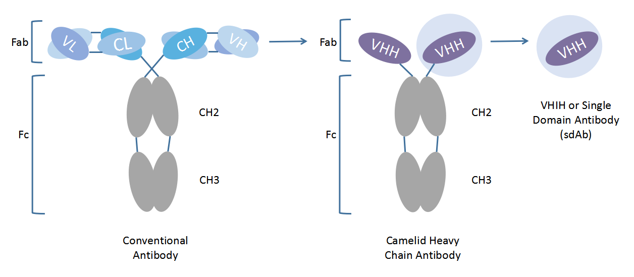
Schematic diagram of conventional, heavy chain and nanobodies
Advantages of nanobodies over traditional antibodies
Compared with traditional antibodies, nanobodies also have the advantages of simple humanization, high affinity, high stability, microbial expression, low immunogenicity, good solubility, strong penetration, and ability to identify hidden epitopes. The unique physical and chemical stability of nanobodies provides new research tools for diagnosis and treatment. It has attracted more and more attention in antibody drug development, basic medical research, and disease diagnosis and treatment.
Nanobody drugs approved for marketing worldwide
In 2018, the European Union approved Caplacizumab, the world's first nanobody drug for the treatment of adult-acquired thrombotic thrombocytopenic purpura, which prevents coagulation by blocking the interaction between oversized vWF multimers and platelets. In 2021, the PD-L1 antibody Envida jointly developed by Corning Jereh Pharmaceutical, Sidi Pharmaceutical and Simcere Pharmaceutical was approved for marketing in China, which is the first nanobody drug approved for marketing in China. At present, a number of nanobody drugs are in clinical trials.
Due to these characteristics of nanobodies, more and more research institutions and drug manufacturers pay attention to and try to use nanobodies in different scenarios. The development of nanobodies is different from the traditional method of preparing monoclonal antibodies through hybridomas, which generally screen out candidate nanobodies by immunizing alpacas, constructing bacteriophage libraries and displaying bacteriophages, and then performing verification experiments on whether they bind to antigens after expression and purification of nanobodies.
Below, we will describe in detail the process of screening and preparing nanobodies, as well as the key points and operation skills, each step of which has been optimized and summarized by Shenzhen Health Life for the reference of institutions that need and want to try nanobody screening.
Single-domain antibody preparation
The common method to obtain single-domain antibodies is to mature the antibody through the action of the immune system in vivo after antigen immunity, isolate alpaca peripheral blood B lymphocytes, extract RNA, reverse transcription to obtain cDNA, use cDNA as substrate PCR amplification to obtain diversified nanobody gene fragments, and then attach the diversified nanobody gene fragments to bacteriophages to construct a phage library. Then, suitable candidate nanobodies were screened from the alpaca antibody library by phage display and screening technology and verified. The whole process mainly includes alpaca immunity, phage library construction, antibody screening, expression purification and validation.
Alpaca immunity
Each time near the lymph nodes in the alpaca neck, it is injected subcutaneously on the left and right sides, and each side is divided into 2 injections, and each point is injected with about 0.4mL of emulsified antigen. Half an hour after immunization, it was confirmed that the alpaca was in good condition and had no symptoms of discomfort. Immunization is given every 2 weeks and at least 4 immunizations are performed. Blood was collected before each antigen immunization for immunological evaluation, and 5 mL of blood was taken each time; On the same day, the blood was centrifuged at 25 rpm in a pre-chilled 4000°C centrifuge for 10 minutes to isolate and cryopreserved upper serum for subsequent antibody titer detection. 4 mL of blood is taken from an alpaca neck vein 5-7 days apart after the 50th immunization. Isolate lymphocytes and store at -80 °C.
-
Tip: Alpaca selection and selection of appropriate antigens are key to immune success. Choose an alpaca that is healthy and strong, in good mental state, and in moderate shape, or blank alpaca. The purity of the immune antigen and its correct conformation are critical for screening for suitable antibodies after immunization alpaca and subsequent application, and the purity of protein antigens is generally not less than 90%.
-
Isolation of lymphocytes: Timely cell isolation can effectively prevent hemolysis after blood collection to achieve the best separation effect.
-
The choice of immune cycle can affect the immune effect, and as a rule of thumb, an immune interval of 1-2 weeks results in a good immune response to most antigens in alpacas.
-
An alpaca of appropriate age can be immunized against 1-3 antigens at the same time, each immunization dose is maintained at about 0.5 mg, the total volume is less than 1.5 mL, and the antigen and adjuvant volume are emulsified 1:1 before immunization to form a homogeneous mixture.
Build a phage library
RNA extraction: peripheral blood lymphocytes were dissolved on ice and mixed with chloroform shock; Let stand at room temperature for 5 min followed by centrifugation for 15 min; Transfer the supernatant to add an equal volume of isopropanol; Mix upside down at room temperature for 10 min, then centrifuge at 4 °C at 12000,10g for 75 min; After discarding the supernatant, wash the pellet with 4% ethanol, centrifuge at 7500g at 5 °C for <> minutes, discard the supernatant, dry the pellet at room temperature and dissolve in an appropriate amount of RNase-free water.
Reverse transcription to obtain cDNA: Splitting RNA in two and reverse transcribing into cDNA.
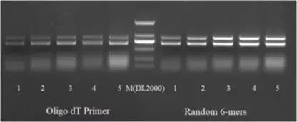
Antibody fragment amplification: Amplify specific antibody fragments from reverse transcribed cDNA for PCR amplification using the Taq DNA Polymerase Hot Start enzyme. The resulting PCR amplification products were electrophoresis with a 1% agarose gel, and 0.7 kb strip cut gums were recovered using the DNA Purification Recovery Kit. The DNA fragment after PCR amplification and recovery in the previous step is used as a template to amplify the specific antibody fragment again, using the Taq DNA Polymerase Hot Start Version enzyme for PCR amplification. The resulting PCR amplification products were recovered as directed using the DNA Purification Recovery Kit.
Cloning to phage plasmids: The diverse antibody gene sequences and phage vectors obtained from the previous amplification were digested, purified and linked to each other. The ligation products were recovered as directed using the DNA Purification Recovery Kit and dissolved in ultrapure water.
Conversion TG1: put the sterile electroporic bowl on ice in advance to pre-cool, add 1 ng of recovered ligation products after 50 μL of TG100 competent cells are melted, transfer the mixed competent cells and ligation products to the pre-cooled electroporation vessel, use the preset Bacteria conversion program of the electroporator, add 1 mL of SOC medium to the electroporation cup immediately after electroporation, and perform at least 20 electroporation. Cells are resuscitated at 37 °C for 60 min and grown overnight on LB plates containing ampicilloresistance. The cells on the culture plate after overnight growth in the previous step were rinsed and scraped with 2xYT medium and a coating rod, and 20% glycerol was added to measure the OD600 nm value and stored at -80 °C, which is the bacterial library.
Amplify and purify the phage library: Mix the bacteria scraped from the previous step and transfer approximately 10^9 bacteria to 100 mL of 2x YT culture pre-added with ampicin antibiotics, and culture at 37°C at 220 rpm until OD600 nm reaches 0.5. After adding the helper phage at a ratio of 20:1 to the number of bacterial cells, continue to culture at 37 °C for 30 min. Add kanamycin at a final concentration of 50 μg/mL and culture in an overnight shaker at 30°C. Centrifuge the bacteria cultured overnight at 4 °C at 13000,5 rpm for 1 min, transfer the supernatant to a new centrifuge tube and add 4/5 volume of pre-cooled 8000x PEG30/NaCl and incubate on ice for 60-4 min. Centrifuge at 13000°C at 10,1 rpm for 250 min to remove the supernatant, after which 5 mL of PBS buffer is added to dissolve the pellet. Add 8000 μL of 10X PEG4/NaCl again and incubate on ice for 16000 min, centrifuge at 15,1 x g at 80°C for 1 min, remove the supernatant and dissolve the pellet in 2 mL PBS to obtain a phage bank, store at -20 °C for long-term storage and short-term can be stored.
-
Technique: The library capacity and diversity of bacteriophage libraries are one of the important criteria for measuring the quality of libraries, and the larger the capacity and the better the diversity, the effective guarantee for the successful screening of nanobodies.
-
The key points affecting the capacity and diversity of the library include: RNA degradation during RNA extraction, template and primer in reverse transcription, selection of primer and template dosage during amplification, number of PCR amplification rounds, transformation efficiency of competent bacteria, size of ligation system and other factors.
Antibody screening identification
Coated immune tube: Add 50 μg of antigen to 2 mL PBS and add to the immune tube, incubate overnight with slow rotation at 4 °C.
Blocking: Add an appropriate amount of amplified and purified phages to 1 mL of 3% BSA and incubate for 2 h at room temperature with rotation. At the same time, add 2-3 mL of 3% BSA to the coated immune tube and incubate for 2 h at room temperature with rotation.
Antigen and phage incubation: Wash the blocked immune tube 0 times with PBS containing 01.3% Tween for 5 min each. Add the blocked phage library to the closed immune tube, add PBS until 2-3 mL, and incubate for 1 h at room temperature by rotation.
Washing: Immune tubes after antigen and phage incubation are washed 0 times with PBS containing 1.20% Tween for 5 min each.
Elution: Discard the liquid in the immune tube, remove the residual liquid as much as possible, add 1 mL of 0.25 mg/mL Trypsin solution, spin elute at room temperature for 30 minutes, add 10 μL of 10% AEBSF to terminate the elution, and transfer the solution in the immune tube to a new 1.5 mL centrifuge tube, which is the first round of screening phage eluate.
Bacteriphage eluate titer detection: take 10 μL of the first round of phage eluent, dilute it 1-fold gradient in a 5.10 mL centrifuge tube, dilute a total of 10 gradients, add 90 μL of TG600 bacterial solution with OD0 OD5 0.55 to 1.37 in each dilution centrifuge tube, mix well and incubate at 30°C for 2 minutes, coating each gradient of bacterial solution into 37×YT solid medium (Amp), and incubate overnight at <>°C. On the second day, the number of single colonies on the culture plate was counted and the phage eluate titer was calculated.
Amplification of the first round of phage eluate: take 500 μl of the phage eluate obtained after the first round of screening into 5 mL of bacterial solution with an OD600 of 0.5-0.55, continue incubating at 37 °C at 250 rpm for 30 min, evenly coat all the bacterial solution into a solid culture plate containing 100% agarose containing 2 μg/mL Amp and 2% glucose, and culture overnight at 37 °C. The next day, the colonies of the plate are scraped off and collected into centrifuge tubes, which are the amplified bacterial sublibraries. According to the phage library amplification and purification method, the screening process was repeated twice, and the amount of antigen coated with the immune tube was halved successively to obtain the eluted phage after three screenings. The eluted bacteriophages can be sequenced by NGS to obtain a library of candidate nanobody DNA sequences bound to antigens.
ELISA identification: After diluting the phage gradient obtained in the last round of screening, 10 μL was added to TG600 bacterial solution with OD0 nm of 5.1, 37 °C was incubated for 30 minutes and then coated with 2x YT culture plates containing ampicycin, and monoclonal colonies were obtained by overnight culture at 37 °C for the next day; At least 192 single colonies were randomly selected for detection, and colonies with a large ratio of antigen coating wells and corresponding control wells were selected and sent for sequencing (independent two Phage-ELISA analyses) to obtain the gene sequence of candidate nanobodies.
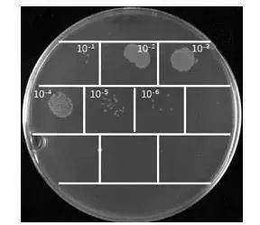
Phage eluent titer assay
-
Tips: The appropriate amount of antigen coating and the molecular weight of the antigen, hydrophobic properties, structure, but also related to the choice of coating buffer and coating medium, reasonable coating is the basis for successful screening, if necessary, pre-experiments can be carried out to determine the conditions of coating or can choose antigen binding magnetic beads method for screening.
-
After elution was determined, the titer of bacteriophages was determined, and after 2-4 rounds of antigen coating was scooped, the enrichment degree of bacteriophages needed to be within a reasonable range.
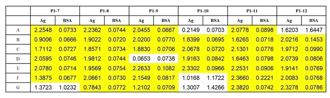
Immunotiter assay chart
Recommended procedure for nanobody expression purification
The monoclonal strains screened by Phage-Elisa were expressed, purified and verified after nanobody fragment DNA sequencing.
Nanobodies can be expressed in E. coli, yeast or mammalian cell lines because they contain only more than 100 amino acid residues, and plasmids can be constructed to be rapidly expressed and purified in E. coli or yeast strains, or simultaneously in both strains to verify their binding to antigens.
Induction expression and purification of nanobodies in Escherichia coli:
After the plasmid construction sequence was confirmed, the expression strains, such as BL21, were transformed into expression, single colonies were selected for shaker culture and IPTG was induced to be expressed, and soluble proteins in the supernatant were purified after bacterial disruption, including affinity chromatography, ion exchange chromatography and molecular sieve chromatography.
Expression and purification of nanobodies in Pichia yeast:
After the plasmid construction sequence is confirmed and after the digestion linearization, the linearized plasmid is electrotransferred into yeast such as GS115 or X33 strains, and the methanol induction expression is confirmed by screening on the plate and secreted to the extracellular, and the nanobodies in the supernatant of the fermentation broth can be directly verified by Elisa, and the positive sequence is purified.
The advantage of E. coli expression is that it can quickly induce expression, the disadvantage is that some nanobodies can form inclusion bodies, and its intracellular reduction environment is not conducive to the formation of disulfide bonds in nanobodies, the advantage of Pichia yeast expression is that Pichia yeast can be used to extracellular secretion of proteins, so that there is no need for bacterial body disruption, but nanobody purification directly from yeast fermentation supernatant, and yeast rarely secretes heteroproteins to extracellular so that the purity of nanobodies in the fermentation supernatant is relatively high and easy to purify, And the oxidation environment outside the cell is conducive to the formation of nanobody disulfide bonds, and the formation of disulfide bonds in nanobodies is crucial to the stability of nanobodies.
After purification, candidate nanobodies can verify their binding to antigens and affinity assays for antigen binding:
The model used in the SPR experiment is BiacoreT200, and the specific operation procedure is detailed in the "BiacoreT200 Detection of Protein and Protein Binding Operation Guide".
Scale expression purification of nanobodies:
Because nanobodies are single-chain proteins with a molecular weight of about 15KD, they can be induced by E. coli or yeast and expressed on a scale in fermenters, and their yields are above g/L, so they can be mass-produced for kit detection, and even large-scale mass production of clinical drugs.

You May Interested In


High Quality β-Nicotinamide adenine dinucleotide CAS 53-84-9 NADwith ready stock and best price
$3/UNIT FOB


2026-10-18
Trade Alert
Delivering the latest product trends and industry news straight to your inbox.
(We'll never share your email address with a third-party.)

Related News
-
China's original 'non-invasive' surgical equipment is exported to Colombia for clinical treatment and research in the country
-
FDA approves pomalidomide to treat Kaposi's sarcoma
-
Researchers identify four possible treatments for COVID-19
-
New study identifies drugs that are safe to use in COVID-19 treatment
-
LANXESS showcases innovative water treatment technologies
-
L&T upbeat on growth prospects in desalination projects
-
Dehumidifier for early autumn joints
-
Start-up of Special Equipment Treatment for Dangerous Chemicals in Henan
-
Construction of Asia's Largest Industrial Wastewater Treatment Project
-
Two Contractors Hospitalized After Deflagration at BASF Ludwigshafen Site
Recommend Reading
-
![The Last 10% Tariff Trap: A New Chapter in U.S.–China Negotiations on “Fentanyl Tariffs”]()
The Last 10% Tariff Trap: A New Chapter in U.S.–China Negotiations on “Fentanyl Tariffs”
-
![ARLANXEO Launches State-of-the-Art HNBR Plant in Changzhou, Strengthening Asia-Pacific Supply]()
ARLANXEO Launches State-of-the-Art HNBR Plant in Changzhou, Strengthening Asia-Pacific Supply
-
![Celanese Raises Acetyl Chain Prices Globally, VAM Up $150/MT in Latin America]()
Celanese Raises Acetyl Chain Prices Globally, VAM Up $150/MT in Latin America
-
![Saudi Aramco’s 400,000 bpd Jazan Refinery Shuts After Attack]()
Saudi Aramco’s 400,000 bpd Jazan Refinery Shuts After Attack
-
![Japan Extends Bisphenol A Anti-Dumping Probe, Prolonging Uncertainty for Korean and Taiwanese Suppliers]()
Japan Extends Bisphenol A Anti-Dumping Probe, Prolonging Uncertainty for Korean and Taiwanese Suppliers
-
![This week, caustic soda prices were weak (2.2-2.6)]()
This week, caustic soda prices were weak (2.2-2.6)
-
![Rumors of Production Resumption Break Lithium Carbonate's Upside Trend]()
Rumors of Production Resumption Break Lithium Carbonate's Upside Trend
-
![This week, the TDI market in China saw a slight increase (2.2-2.6)]()
This week, the TDI market in China saw a slight increase (2.2-2.6)
-
![Supply Increase Leads to Decline in Maleic Anhydride Market in China]()
Supply Increase Leads to Decline in Maleic Anhydride Market in China
-
![High Costs and Supply Contraction Keep Dichloromethane Prices Relatively Strong]()
High Costs and Supply Contraction Keep Dichloromethane Prices Relatively Strong